ELECTRODIAGNOSTIC REFERENCE VALUES FOR ULNAR MOTOR NERVE CONDUCTION RECORDED FROM THE FLEXOR CARPI ULNARIS MUSCLE
DOI:
https://doi.org/10.55374/jseamed.v10.310Keywords:
electrodiagnosis, nerve conduction study, ulnar nerve, reference values, flexor carpi ulnarisAbstract
Background: Ulnar nerve entrapment at the elbow is the second most common compressive neuropathy of the upper extremity. The flexor carpi ulnaris (FCU) muscle may improve diagnostic accuracy in localizing ulnar neuropathy, particularly when distal recording is not feasible because of severe muscle atrophy.
Objective: The present study aimed to establish the electrodiagnostic reference values for the ulnar nerve recorded at the FCU muscle.
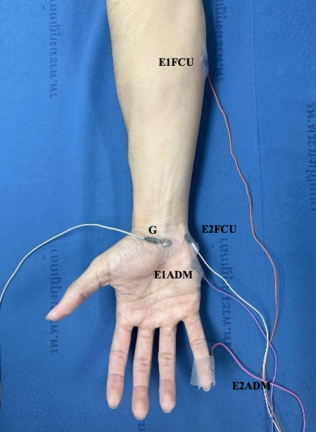
Methods: A total of 73 upper extremities from 44 patients with suspected carpal tunnel syndrome underwent nerve conduction study. For the FCU muscle, the active electrode was placed 10 cm below the medial epicondyle, and the reference electrode was placed at the pisiform bone. The ulnar nerve was stimulated at the wrist, the medial epicondyle, and 10 cm proximal to the medial epicondyle. The recorded parameters included onset latency at the elbow and above the elbow, as well as CMAP amplitude, area, and duration.
Results: The mean DML and CMAP amplitude following stimulation at the medial epicondyle were 2.35 ± 0.35 ms and 8.89 ± 2.84 mV, respectively. The mean NCV across the elbow was 65.12 ± 10.28 m/s. Conclusion: The upper limit of normal for DML was 3.05 ms, and the lower limit of normal (LLN) for CMAP amplitude was 3.21 mV, both derived using the mean ± 2 SD. In contrast, the LLN for NCV across the elbow was 50.51 m/s, based on the 2.5th percentile.
Downloads
Metrics
References
An TW, Evanoff BA, Boyer MI, Osei DA. The prevalence of cubital tunnel syndrome: A cross-sectional study in a U.S. Metropolitan Cohort. J Bone Joint Surg Am 2017; 99: 408-16. DOI: https://doi.org/10.2106/JBJS.15.01162
Osei DA, Groves AP, Bommarito K, Ray WZ. Cubital tunnel syndrome: incidence and demographics in a National Administrative Database. Neurosurgery 2017; 80: 417-20. DOI: https://doi.org/10.1093/neuros/nyw061
Dy CJ, Mackinnon SE. Ulnar neuropathy: evaluation and management. Curr Rev Musculoskelet Med 2016; 9: 178-84. DOI: https://doi.org/10.1007/s12178-016-9327-x
Palmer BA, Hughes TB. Cubital tunnel syndrome. J Hand Surg Am 2010; 35: 153-63. DOI: https://doi.org/10.1016/j.jhsa.2009.11.004
Dumitru D, Zwarts MJ. Chapter 24 - Focal peripheral neuropathies. In: Dumitru D, Amato AA, Zwarts M, editors. Electrodiagnostic Medicine (Second Edition). Boston: Hanley & Belfus; 2002. p. 1043-126. DOI: https://doi.org/10.1016/B978-1-56053-433-4.50032-8
Benecke R, Conrad B. The value of electrophysiological examination of the flexor carpi ulnaris muscle in the diagnosis of ulnar nerve lesions at the elbow. J Neurol 1980; 223: 207-17. DOI: https://doi.org/10.1007/BF00313185
Felsenthal G, Brockman PS, Mondell DL, Hilton EB. Proximal forearm ulnar nerve conduction techniques. Arch Phys Med Rehabil 1986; 67: 440-4.
Awang MS, Abdullah JM, Abdullah MR, Tahir A, Tharakan J, Prasad A, et al. Nerve conduction study of healthy Asian Malays: the influence of age on median, ulnar, and sural nerves. Med Sci Monit 2007; 13: CR330-2.
Fong SY, Goh KJ, Shahrizaila N, Wong KT, Tan CT. Effects of demographic and physical factors on nerve conduction study values of healthy subjects in a multi-ethnic Asian population. Muscle Nerve 2016; 54: 244-8. DOI: https://doi.org/10.1002/mus.25029
Ginanneschi F, Curti S, Marinelli F, Nft AA, Cioncoloni D, Mattioli S, et al. Reference values for distal motor conduction of the tibial nerve: Effects of demographic and anthropometric measures. Muscle Nerve 2020; 62: 219-25. DOI: https://doi.org/10.1002/mus.26908
Lafratta CW, Smith OH. A study of the relationship of motor nerve conduction velocity in the adult to age, sex, and handedness. Arch Phys Med Rehabil 1964; 45: 407-12.
Egbuchulem KI. The basics of sample size estimation: an editor’s review. Ann Ib Postgrad Med 2023; 21: 5-10.
Chen S, Andary M, Buschbacher R, Del Toro D, Smith B, So Y, et al. Electrodiagnostic reference values for upper and lower limb nerve conduction studies in adult populations. Muscle Nerve 2016; 54: 371-7. DOI: https://doi.org/10.1002/mus.25203
Lee WS, Yang H-J, Park SB, Son YJ, Hong N, Lee SH. Location of ulnar nerve branches to the flexor carpi ulnaris during surgery for cubital tunnel syndrome. J Korean Neurosurg Soc 2023; 66: 90-4. DOI: https://doi.org/10.3340/jkns.2022.0128
Downloads
Published
How to Cite
Issue
Section
License
Copyright (c) 2026 Journal of Southeast Asian Medical Research

This work is licensed under a Creative Commons Attribution-NonCommercial-NoDerivatives 4.0 International License.
The Journal of Southeast Asian Medical Research will hold the copyright to all published articles. The publisher's production department handles copyright forms once a manuscript is accepted and scheduled for publication.